Multiplex immunofluorescence (MxIF) is a recently developed imaging platform whereby a large number of cellular and histological markers can be investigated on a single tissue section.
Standard immuno-detection techniques, such as immunohistochemistry (IHC) and immunocytochemistry (ICC), only allow for 1-5 markers (6 for multi-spectral microscopy) to be studied simultaneously per cut section of tissue. Probing for more markers requires that multiple serial sections be cut from the same tissue block, which could deplete rare and precious samples. Moreover, the resulting data gleaned from each serial section cannot be directly related to the other sections, as each of the individual cuts do not contain the exact same cells and possible tissue micro environment as the others.
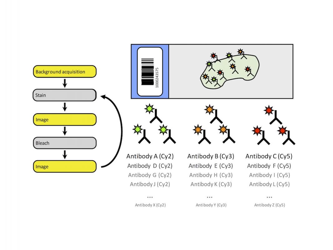
This fundamental issue of direct marker comparison is addressed by MxIF. This method relies on repeated rounds of staining, imaging, dye inactivation, and background subtraction in order to layer multiple markers onto the same regions of interest, thus enabling researchers to gain high-dimensional data on a single cut of tissue. Additionally, tissue level and single-cell segmentation can be accomplished by localizing multiple cellular markers, enabling subcellular quantification of antibody staining as well as measurement of cell size, shape, and position.
Over the last four years, members of the Epithelial Biology Center (where the DHSR is housed), in conjunction with GE Healthcare, have been part of an NIH Common Fund grant with the goal of developing a system for multidimensional immunofluorescence. The DHSR houses one of three prototype GE Cell-DIVE™ MxIF platforms developed from that collaboration.