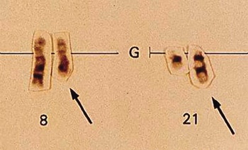
A fusion protein resulting from a faulty DNA exchange plays a key role in the development of acute leukemia in humans, Vanderbilt researchers reported recently. The protein, called AML1-ETO, disarms a tumor suppressor and allows circumvention of a powerful tumor checkpoint, freeing cells to experience uncontrolled growth and an extended lifespan.
A reciprocal exchange, or translocation, of genetic material between two chromosomes is a mistake that can occur as a cell duplicates its DNA, just before it divides. The chromosome translocation – in which a piece of one chromosome breaks off and fuses to a second chromosome – is passed along to successive generations of cells.
The t(8;21) translocation, where DNA is exchanged between chromosomes 8 and 21, occurs in about 15 percent of all cases of acute myeloid leukemia, a cancer of the blood in which certain white blood cells proliferate, crowding out normal cells in bone marrow and infiltrating organs such as the liver or spleen. The translocation juxtaposes two genes that are not typically neighbors. Fusion of the genes produces the unique protein, AML1-ETO.
Although many types of leukemia have been associated with chromosomal translocations, the means by which a resulting fusion protein can cause cellular growth to go awry is not well understood. In the July issue of Nature Medicine, Scott W. Hiebert, Ph.D., professor of Biochemistry, and graduate student Bryan Linggi, report that AML1-ETO represses transcription of a known tumor suppressor, p14ARF. Their studies suggest that loss of p14ARF allows the cell to bypass the formidable p53 tumor checkpoint.
“Basically, we’ve figured out what the fusion protein is, what it does, and how it does it,” Hiebert said. “The translocation takes two parts – two transcription factors – that fuse to make a new transcription factor, which turns off, or inactivates, a tumor suppressor. It’s just a different way to turn off a suppressor.”
There are two opposing factors at work in cancer, according to Hiebert. Oncogenes are genes that cause cancer by increasing cell proliferation – that is, abnormal growth. Tumor suppressors put limits on growth – they are the brake. Many of them act as checkpoints, he said. So if an oncogene is turned on, or activated, in a cell, growth could be held in check by one or more of the suppressors.
“If you lose those checkpoints,” Hiebert said, “cell growth will take off. Usually a cancer results from a combination of these events.”
The suppressor p14ARF has been described as the major oncogene checkpoint in humans. And it engages another tumor suppressor called p53, which is mutated in over half of all cancers. If an oncogene is activated, then p14ARF and p53 act as checks. If either p14ARF or p53 is lost, the cells continue to grow unchecked.
What happens if p14ARF is lost, but p53 is intact? Although p53 inactivation is necessary for the development of most cancers, it is rarely mutated in human acute leukemia. Yet it appears that loss of p14ARF impairs p53-mediated cell growth arrest and suicide. Linggi, who is a fifth year graduate student working in Hiebert’s lab, said it is because p53 is implicated in so many cancers that it was important to iron out how the two suppressors are related.
“So it all actually fit together pretty nice in being able to explain how you can have cancer develop and still have expression of p53, with no obvious mutation or deletion,” he said. “We are proposing that suppression of p14ARF by this fusion protein makes that happen.”
Next, the lab will look at other translocations that disrupt AML1 function (associated with 25 percent of all leukemias) to see if they also deactivate the p14ARF tumor suppressor.
The work reported by Hiebert and Linggi was supported by grants from the NIH, the National Cancer Institute, and the Vanderbilt-Ingram Cancer Center.


