Welcome to the Vanderbilt Center for Bone Biology

Investigating the development of the musculoskeletal system as well as diseases and conditions that negatively affect i) musculoskeletal tissue and ii) its ability to heal and regenerate, VCBB investigators aim to develop new treatment strategies and diagnostic tools that can improve the quality of life for patients with compromised bone, articular cartilage, and ligaments due to aging, trauma, infection and disease.

Jeffry S. Nyman, PhD, Director
Our Research

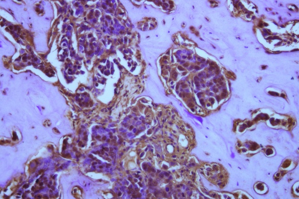
  • Human MCF7 breast cancer cells lie dormant in the bone marrow and stain for pimonidazole (a marker for hypoxic, or low oxygen, conditions) along the tumor-bone interface. Work in the Johnson laboratory suggests that very low oxygen levels may promote breast cancer cells to exit dormancy and colonize the bone marrow.
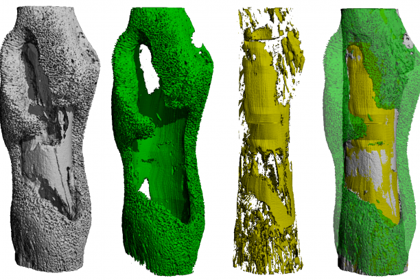
  • Quantitative imaging analysis of pathologic bone remodeling during S. aureus osteomyelitis from Dr. Jim Cassat's laboratory. A 3D rendering of the infected femur by microCT is depicted (left), and calculations of new bone formation and cortical bone destruction are illustrated in green and yellow, respectively.
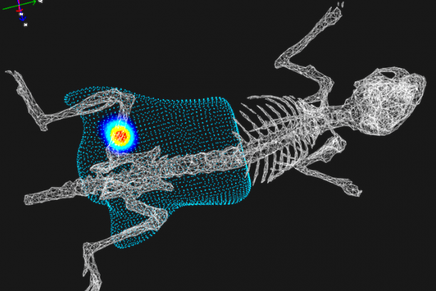
  • 3D bioluminescent imaging reveals a hypoxia-responsive S. aureus promoter in vivo during osteomyelitis, from Dr. Jim Cassat's laboratory.
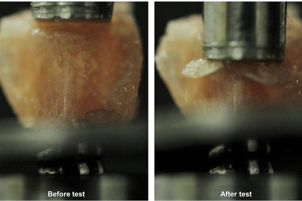
  • Axial compression test of a mouse lumbar vertebra from Dr. Nyman’s lab. Such tests are used to determine the effect of treatment, disease, or gene modification on whole bone strength.
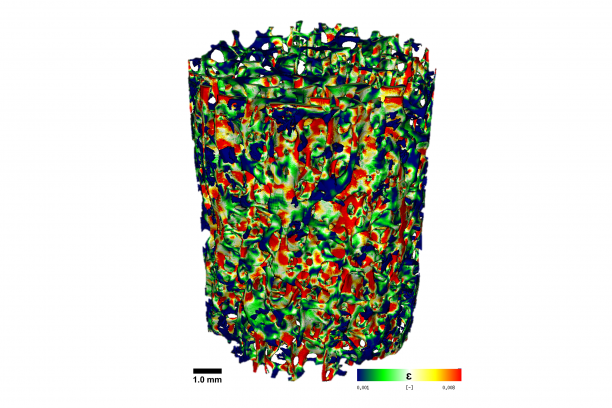
  • Strain distribution for a human trabecular bone core subjected to axial compression as determined micro-CT-derived finite element analysis from Dr. Jeffry Nyman's laboratory. Such calculations identify weak trabeculae (red).
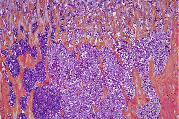
  • MDA-MB-231 human breast cancer cells in the bone marrow of a mouse treated with zoledronic acid, from Dr. Julie Sterling's laboratory.