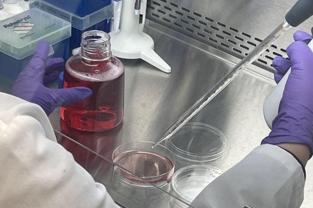
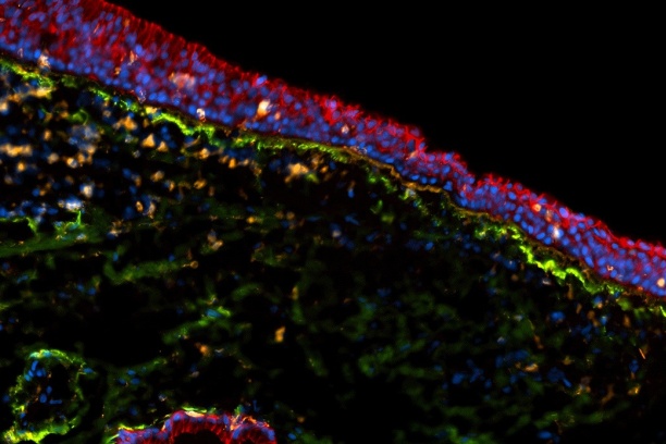
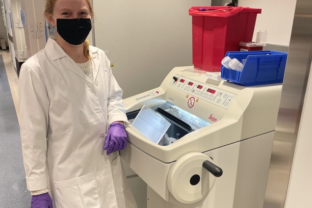
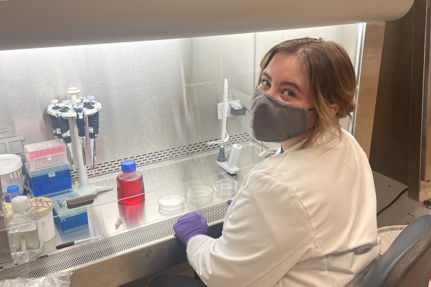
Directed by Dr. Emily Kimball, the Voice Biology Lab focuses on the biological and physiological basis for vocal fold health and pathology. We are particularly interested in understanding the specific cellular and molecular mechanisms by which the vocal folds maintain their ability to vibrate freely. Along with this goal, we also seek to understand the circumstances that ultimately lead to the development of vocal fold lesions like nodules and polyps, which cause disordered voice production.
Our work is divided across a few ideas:
- What keeps the vocal folds healthy in people who don't have voice disorders but use their voice a lot? What is happening in the tissue in these cases?
- How can we better predict when someone is at greater risk of developing lesions? What behavioral and environmental factors actually matter?
- How can we better align clinicians in the way we talk about lesions and voice pathology? Out goal is to communicate better about our patients and draw more specific insight from their treatment outcomes through validated protocols and measures.
It is our hope that the work done in this laboratory can have a positive impact on the clinical care for patients with voice disorders.
