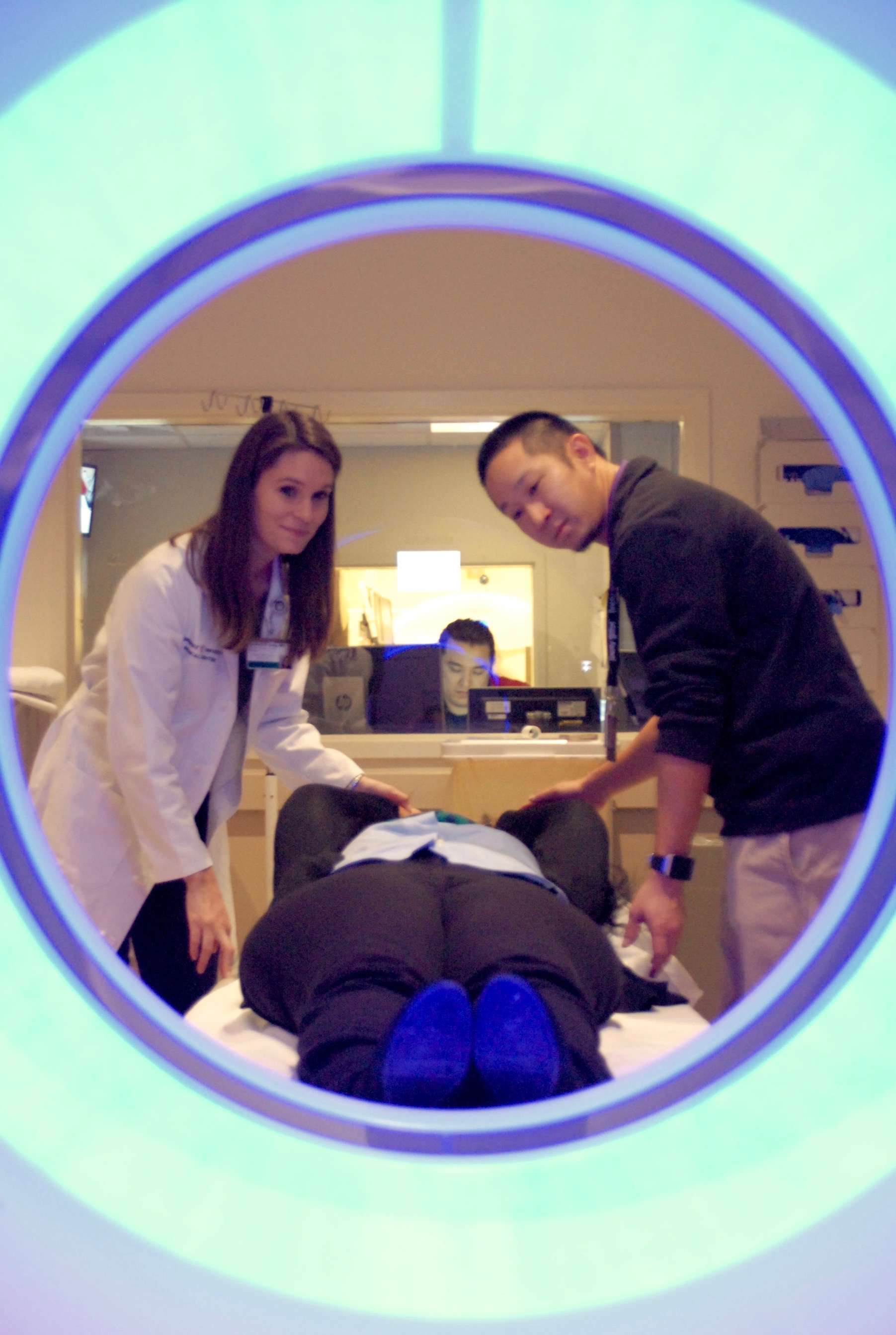
 One of the Department of Radiology’s 11 sub-specialty sections, Body Imaging works closely and efficiently with departments across Vanderbilt University Medical Center to provide image interpretation in computed tomography (CT), magnetic resonance imaging (MRI), ultrasound and fluoroscopy, as well as image-guided interventional procedures.
In an effort to enhance workflow efficiency, as well as visibility across the Medical Center, recently appointed section chief Geoffrey Wile, M.D., Associate Professor of Clinical Radiology and Radiological Sciences, says there will be an even larger emphasis placed on achieving pillar goals in the areas of research, education and patient care this year.
“We’ll accomplish this through a balanced team approach, and by practicing effective communication between groups in on- and off-site imaging locations,” said Dr. Wile. “This will result in more seamless clinical work, including the potential to provide more advanced levels of subspecialty image interpretation and increased visibility in patient engagement.”
Filip Banovac, M.D., has actively been working with the Interventional Radiology section to streamline service provided for procedures, which has resulted in increased cooperation amongst sections, as well as an improved patient and provider experience.
Discussions around new service lines in CT and the introduction of new contrast agents for ultrasound and MRI have also taken place.
In addition to improvements in the clinical practice, there will be a continued focus on career advancement and faculty development, with an increased emphasis on teaching and research opportunities for section members.
“Former section chief Ron Arildsen, M.D., set a department-wide standard for professionalism and career success that I’m committed to upholding with his guidance,” said Dr. Wile.
With the addition of new faculty members Krupa Patel-Lippmann, M.D., and Michael Fleming, M.D., this past year, the section continues to grow and gain members with various clinical experience and research interests.
In recent months, section members have also achieved department- and VUMC-wide recognition through the development and implementation of various teaching and research initiatives. Lori Deitte, M.D., partnered with faculty in Anesthesiology to develop a mobile application, QuizTime, for medical student education. Asma Ahmad, M.D., hosted a hands-on ultrasound procedure session in the cadaver lab for resident education. Rick Abramson, M.D., became a site principle investigator for a multi-site NIH (Quantitative Imaging Network) trial of a software platform for evaluating novel imaging biomarkers. And lastly, Sandeep Arora, M.B.B.S., co-led a team of researchers that investigated new prostate cancer therapy in the TULSA-PRO Ablation Clinical Trial.
“Opportunities such as these are beneficial not only to faculty in the advancement of their careers, but also for patients, trainees and the department as a whole in its effort to cultivate leaders in radiology,” added Dr. Wile.
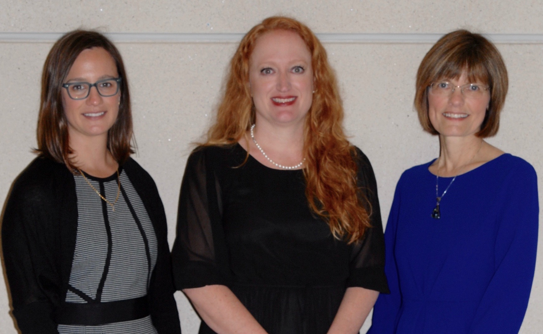
Lori Deitte, M.D. (right), Leslie Fowler, M.D. (center), and Meaghan Magarik, M.D., Ph.D. (left), collaborated on the QuizTime application.
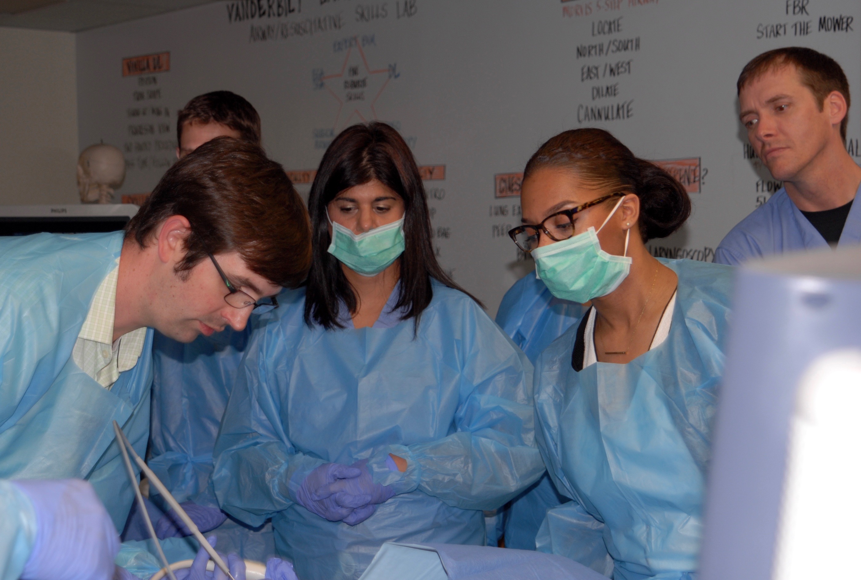
Asma Ahmad, M.D., leads hands-on ultrasound procedure session with radiology residents in the VUMC cadaver lab.