Urinary Microbiology
This research focuses on Urinary Microbiology and Microbiome and is a collaboration between Dr. Reynolds and microbiologists at VUMC.

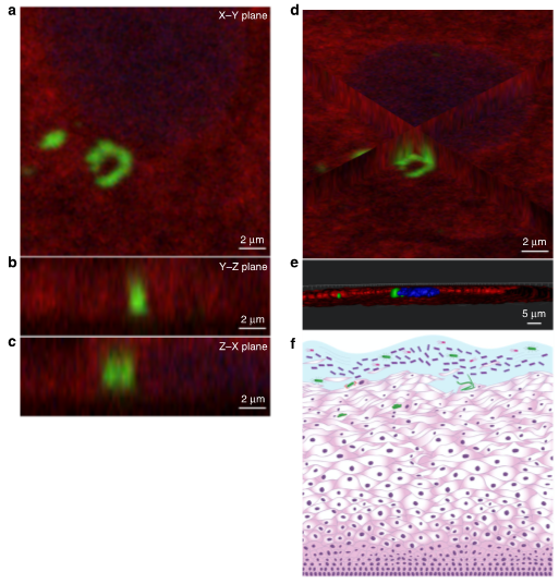
Fig. 4 UPEC invades the VECs of women with a history of rUTIs. For this pilot study, immunofluorescence microscopy was used to search for E. coli within VEC collected by vaginal swabbing of four women with a history of rUTIs. a–d For immunofluorescence, samples were stained with an α-E. coli (green), cytokeratin 13 (red), and α-uroplakin III (yellow) and ToPro-3 (blue). a The XY-plane image from a Z-stack series of a VEC with E. coli surrounded by the VEC-specific cytokeratin 13. Ortho views of b YZ-plane and c XZ-plane reconstructed from a Z-stack series images showing E. coli surrounded by cytokeratin 13 within a VEC near the nucleus. d Ortho-view of images mid-way through the VEC demonstrating the internal localization of invading E. coli on all three dimensions. e Surface reconstructions of E. coli, VEC cytokeratin 13, uroplakin III, and nucleus that shows that the nucleus and E. coli are within the cytokeratin 13 of the VEC. Imaris software was used for image analysis. f Illustration depicting invasion of the vaginal epithelium by UPEC (green bacteria). From Brannon et al, Nature Communications, 2020.

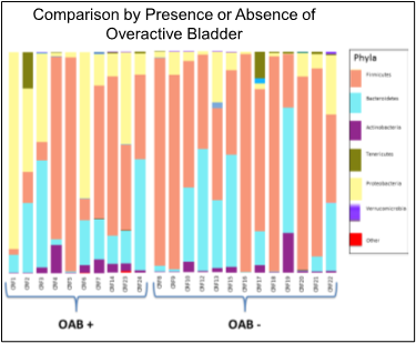
Figure. Pilot project data comparing genitourinary microbiome in women with and without OAB. From Cohn et al, The Journal of Urology, 2017.
Selected Publications
Brannon JR, Dunigan TL, Beebout CJ, Ross T, Wiebe MA, Reynolds WS, Hadjifrangiskou M (2020). Invasion of Vaginal Epithelial Cells by Uropathogenic Escherichia Coli. Nature Communications; 11(1): 2803. PMID: 32499566.
Cohn J, Brown ET, Guo Y, et al. The Relationship Between the Urinary Microbiome and Central Sensitization in Women with Overactive Bladder. The Journal of Urology. 2017;197(4S):e398-399.
