-
Well-designed wet lab space aids safe handling of biological tissue samples, their preparation and storage while designing research protocols and experiments. It is equipped with several ultra-low temperature freezers and various bone cutting and machining tools, as shown below:
Bone cutting equipment
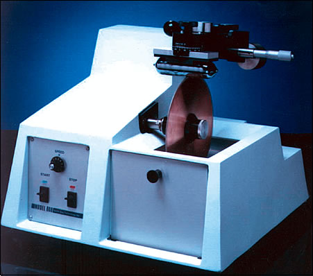
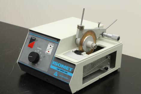
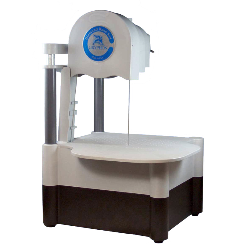
- Osteotomy saws
- South Bay Tech inc., Model: 660 - diamond blade, variable speed, water irrigated.
- Buehler ISOMET, Model: 111280160 - diamond blade, low-speed, water irrigated.
- Gryphon USA inc., Model: C40 - diamond bandsaw, water irrigated.
- Bandsaws: SKIL inc. Model 3385 bench-top saw, Microlux variable speed band saw
- Drill press (RIDGID power tools, Model: DP15501)
- Lathe (JET belt driven bench Model: BD-920W)
- Custom milling-fixtures to make uniform shaped samples out of rough bone cuts
Freezer storage space
- -20°C chest freezer (Model: 55700-385)
- 4°C /-20°C upright combo freezer (Model: 10LCEETSA)
- -86°C Ultra-low temperature upright freezer (Model: ULT 2586-6-A46)
- -80°C Ultra-low temperature upright freezer (Model: IU2386A)
- Osteotomy saws
-
MTS Bionix™ 858 Axial Torsional Test system
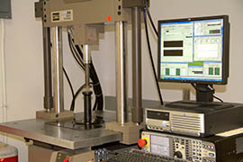
This full size, floor mounted servohydraulic test frame can be used to perform uni- or bi-axial mechanical tests on material samples, implants or specimens using load or displacement control. It can execute biomechanical tests in compression, tension, flexure, and torsion thereby simulating a variety of physiologic forces and motions. The test system is integrated with state-of-the-art digital controls (FlexTest® SE), powerful MultiPurpose Testware (MPT) application software and a host of accessory options (attachment fixtures, load cells, extensometer, etc.) that can be configured to fit a wide variety of testing needs.
Instron DynaMight™ 8800 Servohydraulic Test system
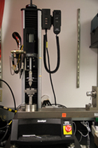
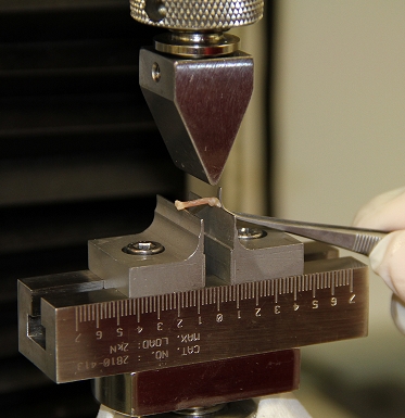
This bench-top, low-capacity servohydraulic test system is capable of performing a wide variety of low-cycle and high-cycle fatigue tests, crack propagation and fracture toughness experiments, as well as other quasi-static tests such as 3-pt, 4-pt bending in load or displacement control modes. The system, combined with advanced features of the 8800 digital controller, FastTrack™ Console software, and WaveMaker™ Editor programming software, and other appropriate accessories (grips, clamps, compression platen, load/torsion cells, extensometer, etc., is ideal for running a variety of static tensile, compression, flexure, peel, tear and friction tests.
Reference Point Indentation system (BioDent, Activelife inc.)
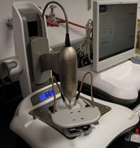
 BioDent is a novel biomechanical instrument used for measuring bone tissue mechanical properties using the principle of cyclic reference point indentation (cRPI). The RPI instrument performs bone microindentation testing to obtain direct measurement of bone tissue strength and is suitable for both in-vitro and in-vivo use. It uses a probe assembly consisting of a reference probe that penetrates the soft tissue above bone and a test probe that is actuated into and out of the bone using a force control algorithm. The measurement system alongside records the dynamics of the test probe such as Indentation distance increase, Creep, loading-unloading slope, etc. to help characterize the material properties in turn.
-
Our research techniques also use several state-of-art imaging tools housed in the Vanderbilt University Institute of Imaging Science’s (VUIIS) Center for Small Animal Imaging (CSAI), and Vanderbilt Biophotonics Center. These collaborations give a more directed approach in tackling the various research questions from an multi-modal imaging standpoint.
Micro Computed Tomography (MicroCT)
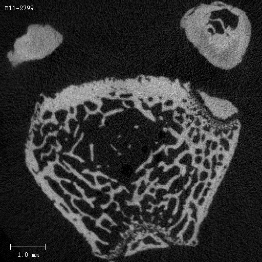
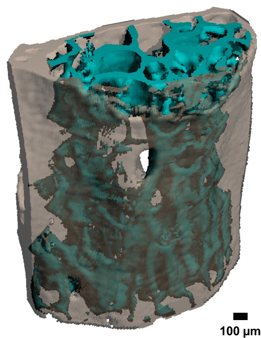 We have both has ex vivo and in vivo imaging capabilities of bone, soft tissues, synthetic materials, and small animals via a host of X-ray CT scanners, Scanco Medical microCT40, microCT50, vivaCT80, and Xtreme CT II HR-pQCT, designed for ultrahigh-resolution scanning to quantify microstructure and architecture. The polychromatic X-ray tube sources operate between 45 and 90 kVp. The scanners can accommodate samples up to 90 mm in diameter and 80 mm in height and with the optimal acquisition settings we can achieve an isotropic voxel resolution as low as 0.5µm. The systems are equipped with dedicated software for image reconstruction, visualization, and analysis. The analysis software includes a full suite of tools for bone morphometry measurements.
 Raman Spectroscopy
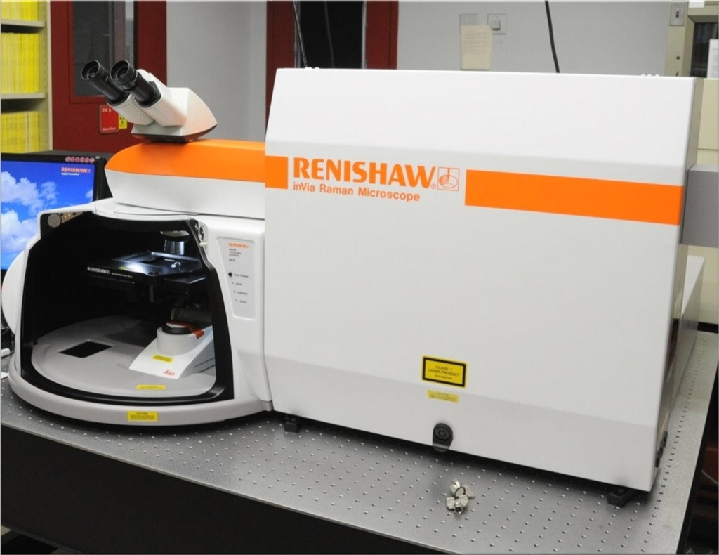
The Vanderbilt Biophotonics Center (Keck FEL Bldg) are focused on the applications of light in medicine and biology. Our research primarily uses the (i) bench-top Raman confocal microscope system (Renishaw inc., Model: inVia) equipped with 785 and 830nm laser source, (ii) combined Raman-OCT system, and (iii) various hand-held imaging probes for SORS technique to assess bone matrix quality and composition.
1H Nuclear Magnetic Resonance (NMR) spectroscopy
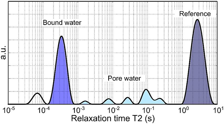 VUIIS houses a 4.7T, 31-cm bore Varian DirectDrive NMR scanner. It is a fully broad-banded imaging/spectroscopy system equipped with actively shielded gradients, two independent transmit and one independent receive channels to quantify water fractions in small bone samples.
High resolution digital SLR camera
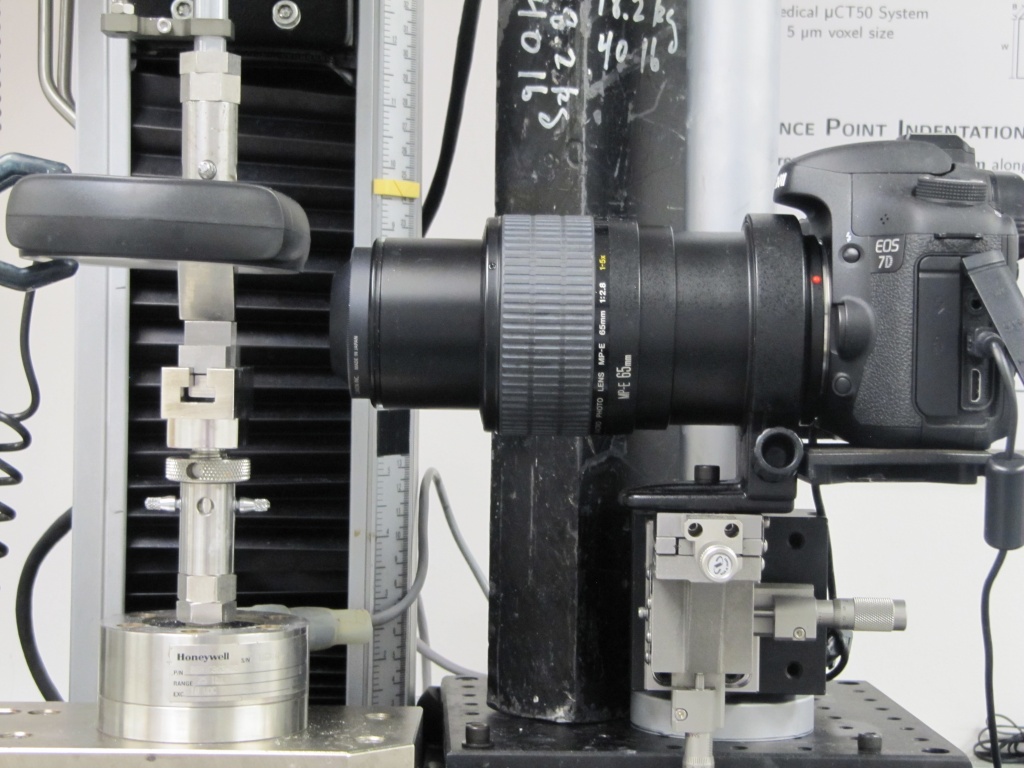
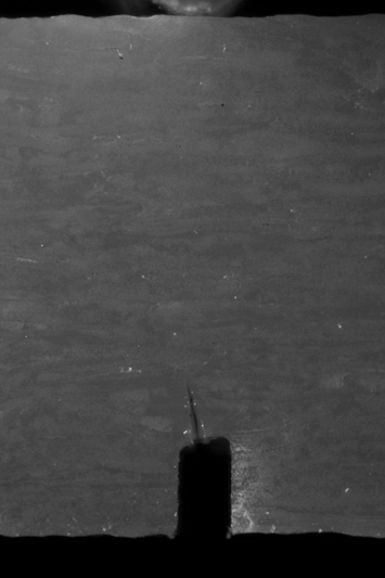
The EOS 7D is a high-performance, digital SLR camera with a fine-detail CMOS sensor capable of 18MP resolution. It also features a powerful Dual DIGIC4 image processor offering a wide range of functionalities. We have multiple accessories such as, LED ring lights, Flash lights, macro lens, trigger timers, holding fixtures etc. to aid a variety of shooting modes.
-
- Matlab (Mathworks inc.) r2017
- STATA statistical software
- GraphPad Prism v6.0 scientific graphing and statistical software
- SolidWorks 2012 3D Computer Aided Design (CAD) support
- Image visualization tools: ImageJ, MeshLab, Dragonfly 3.1
-
- Collagen crosslink analysis by HPLC
- Finite Element Method
- Histomorphometry